Skeletal scintigraphy
Gentle examination of the entire skeleton
What is skeletal scintigraphy?
Skeletal scintigraphy is one of the most common examinations in nuclear medicine. It allows doctors to see changes in bones before they appear on an X-ray or CT scan.
A radioactive substance accumulates where the bone is undergoing change – for example, due to inflammation, injury or cancer.
Why is the examination performed?
- To detect bone metastases in cancer patients.
- When bone inflammation (osteomyelitis) is suspected.
- For complications after surgery and joint implants.
- To examine fractures that are not visible on X-rays.
- For rheumatic and degenerative diseases (arthrosis, arthritis).
- To detect metabolic bone diseases (e.g. Paget's disease).
How is the examination performed?
- A small dose of radioactive substance will be administered into your vein, which will be stored in your bones.
- This is followed by a break (2–3 hours) during which the substance "works". We recommend drinking plenty of fluids.
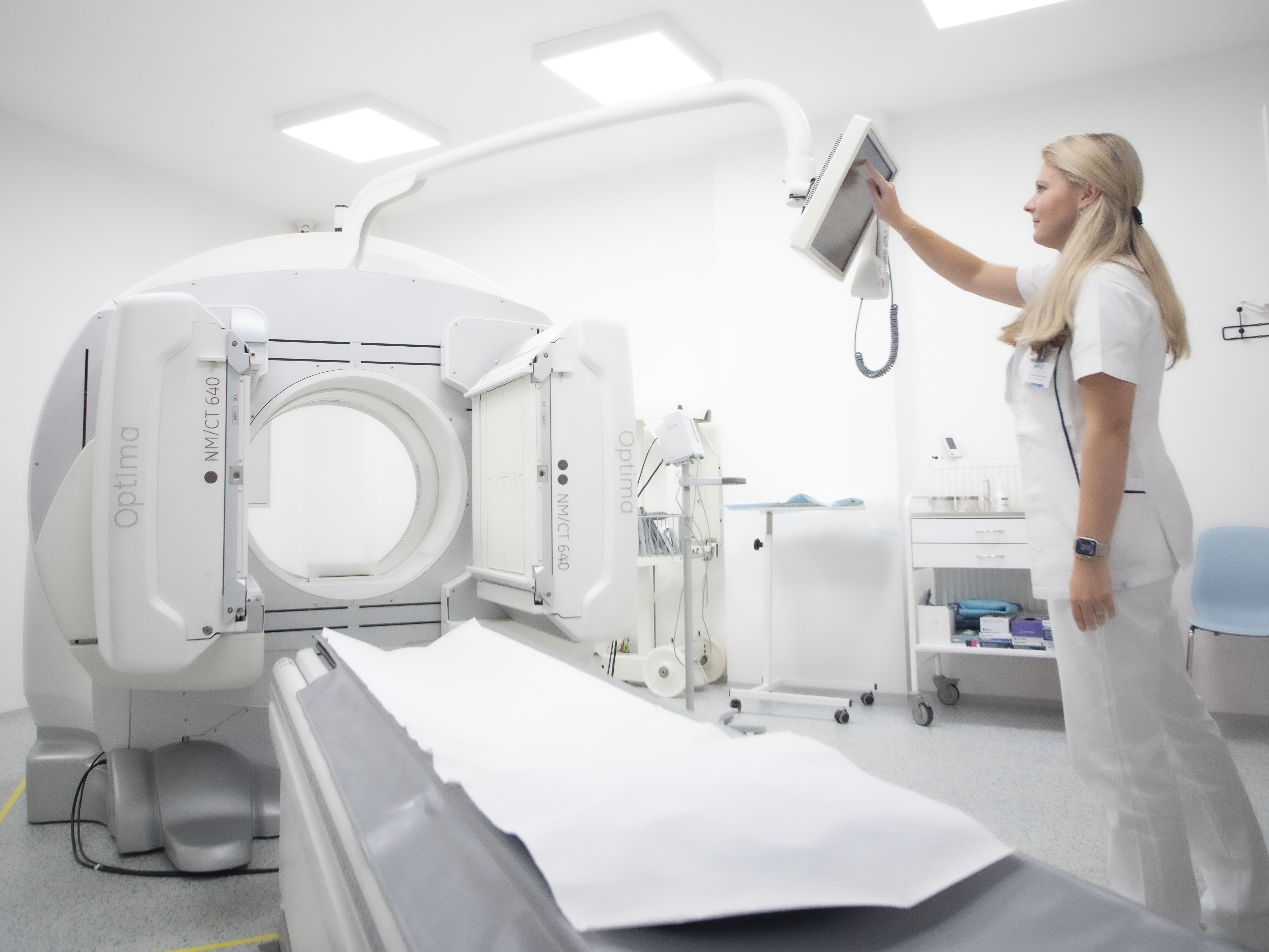
- You will then lie down on the examination table and a special camera will take an image of your entire body.
- The imaging itself takes about 30–60 minutes.
The examination is painless and safe.
How to prepare?
- Drink plenty of fluids on the day of the examination.
- Always empty your bladder before the scan.
- No special diet or discontinuation of regular medication is necessary.
Is the examination safe?
Yes. The radiation exposure is low, less than that of a standard full-body CT scan. The examination is performed routinely and has been used for a long time.
Why choose us?
- Fast and reliable results
- Modern equipment (SPECT/CT) for accurate diagnosis
- Individual approach and experienced team
- We cooperate with all health insurance companies
