Bone marrow scintigraphy
A practical guide for referring physicians
 Principle and method
Bone marrow scintigraphy is a specialised radionuclide examination aimed at assessing the distribution and function of the haematopoietic bone marrow and distinguishing normal haematopoietic activity from pathological infiltrates or displacement of the marrow outside its physiological spaces.
Two basic radiopharmaceuticals are used:
- 99mTc-nanocolloid – colloidal particles captured by the reticuloendothelial system, especially in the bone marrow, liver and spleen. It shows the distribution of phagocytic bone marrow
- Labelled leukocytes – show the distribution of haematopoietic bone marrow
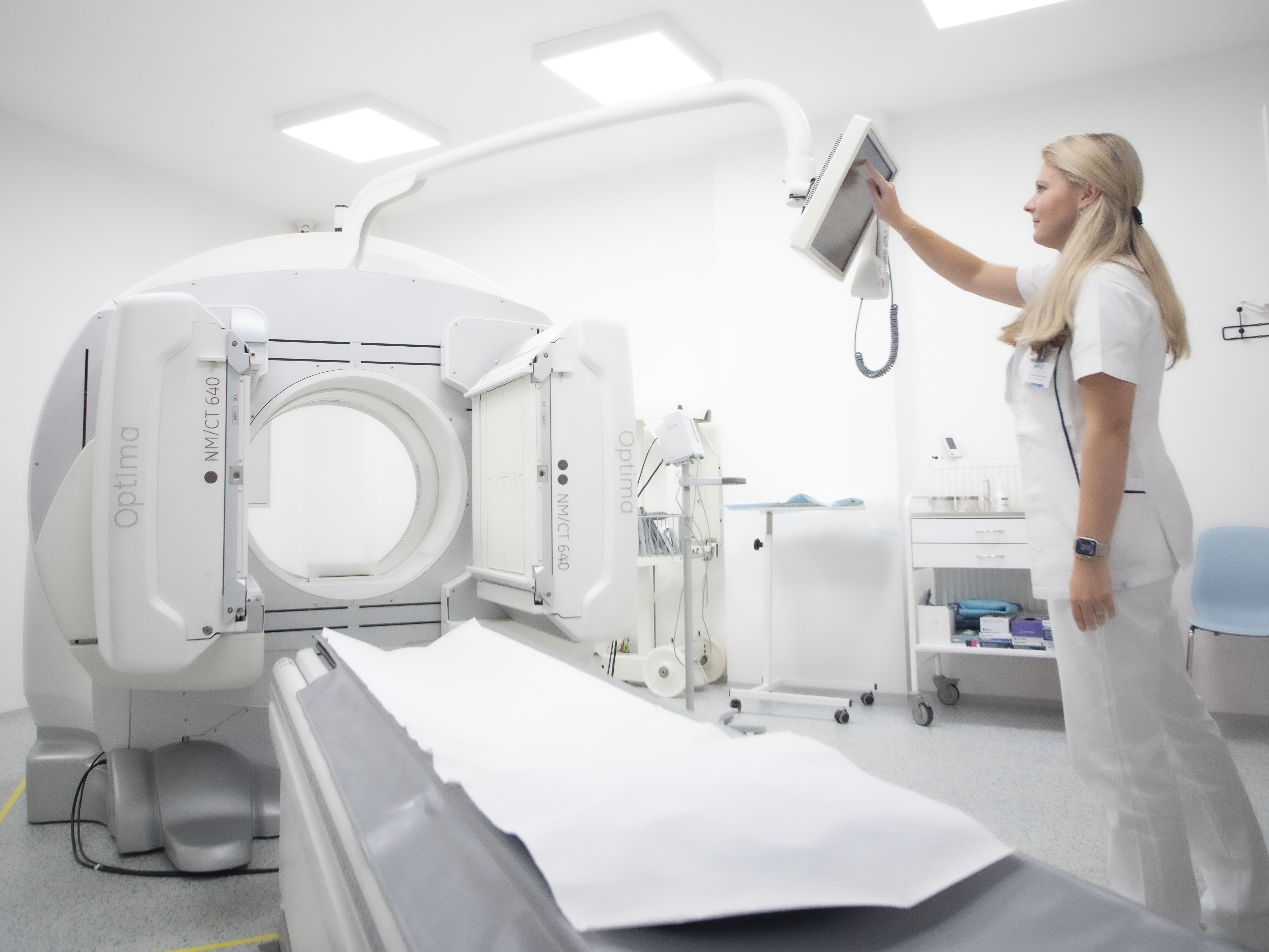
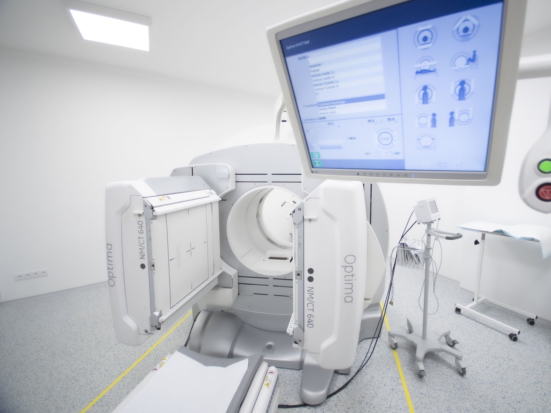
The examination is performed as whole-body planar scintigraphy, supplemented by SPECT/CT for more accurate localisation of pathological foci.
Main clinical indications
- Haematological malignancies
- Acute and chronic leukaemia – assessment of the extent of bone marrow infiltration
- Lymphomas – assessment of bone marrow involvement as part of staging
- multiple myeloma – detection of active foci, differentiation from simple osteolytic changes.
- Myeloproliferative and myelodysplastic syndromes
- detection of extramedullary haematopoiesis,
- mapping of active and inactive areas of the bone marrow.
- Oncological indications
- evidence of bone marrow infiltration by solid tumours (neuroblastoma, carcinomas),
- distinguishing bone metastases from active haematopoietic foci (especially in children and young patients).
- Post-therapeutic monitoring
- after chemotherapy and radiotherapy – assessment of haematopoietic regeneration,
- after bone marrow or peripheral stem cell transplantation – mapping of haematopoietic tissue repopulation.
- Benign conditions
- differential diagnosis of aplastic anaemia,
- assessment of the extent and distribution of bone marrow in haematological diseases of unclear aetiology.
Interpretation
- Normal findings: symmetrical distribution of activity in the axial skeleton (spine, sternum, ribs, pelvis, proximal femurs and humeri). In children, physiological bone marrow activity is more extensive and also includes long bones.
- Pathological findings:
- accumulation defects – corresponding to tumour infiltration, fibrotic remodelling or bone marrow damage,
- extramedullary haematopoiesis – accumulation outside the bone marrow (liver, spleen, paravertebral foci),
- asymmetry – suspected focal infiltration or tumour activity.
Practical information for the referring physician
- Patient preparation: no special diet or discontinuation of medication is necessary.
- Examination procedure:
- i.v. administration of radiopharmaceutical,
- imaging after 30–60 minutes and 2–4 hours
- whole-body examination, supplemented with SPECT/CT as indicated.
- Examination duration: 1–2 hours
- Radiation exposure: 2–5 mSv depending on the type of radiopharmaceutical.
- Contraindications: pregnancy; otherwise no absolute contraindications.
Summary for practice
Bone marrow scintigraphy is:
- an important method for evaluating haematopoietic activity and marrow infiltration,
- useful in haematology (leukaemia, lymphomas, myeloma, myeloproliferative syndromes),
- beneficial in oncology (distinguishing metastases from active haematopoiesis, follow-up after therapy),
- a valuable supplement to MRI and PET/CT, providing unique functional information.
Thanks to its ability to assess the activity and distribution of haematopoietic tissue, this method remains a key tool, particularly in haematology, oncology and transplant medicine.
