Assessment of sympathetic innervation of the heart
Practical guide for referring physicians
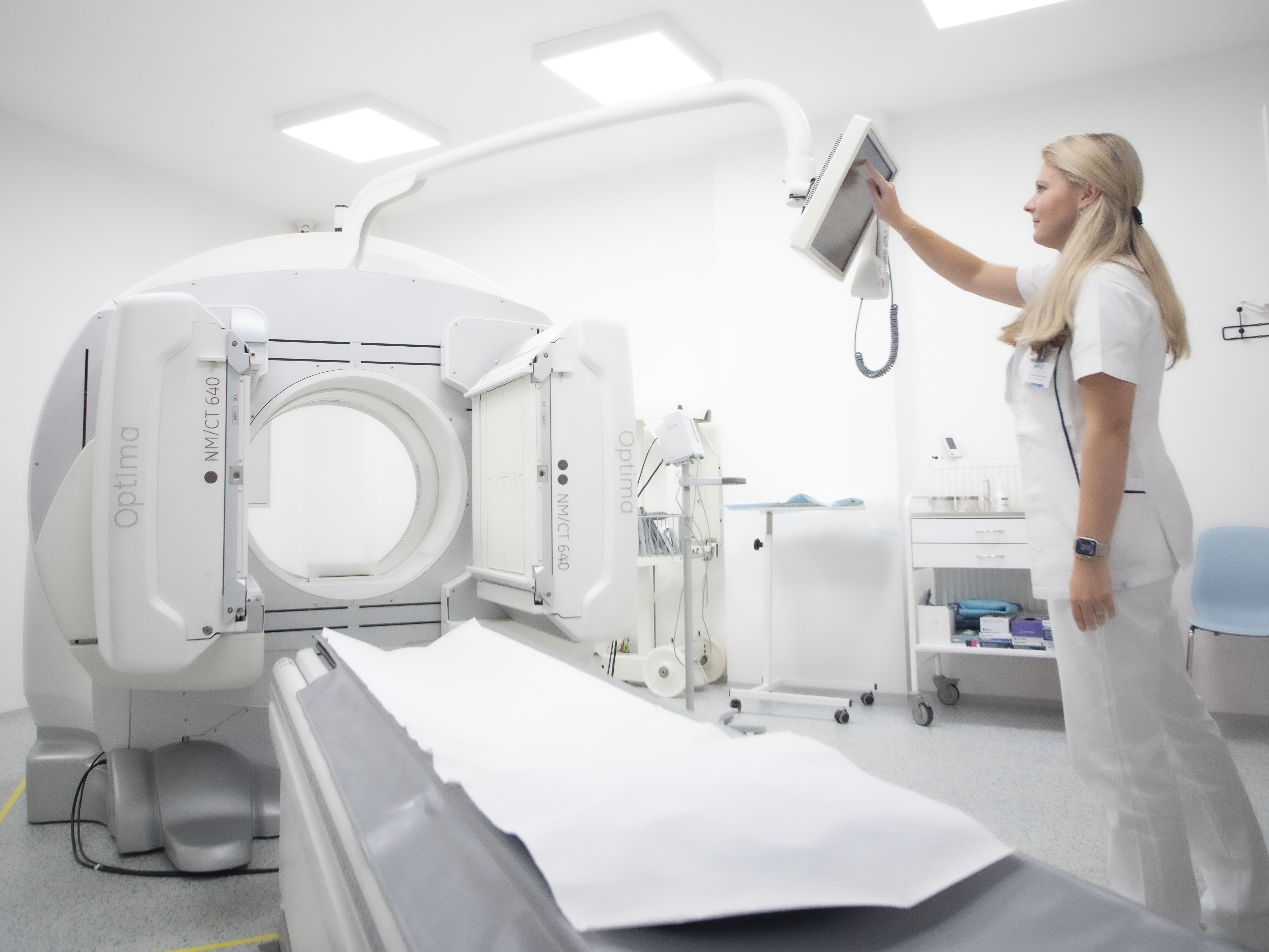
1. Principle and method
123I-metaiodobenzylguanidine (mIBG) is a synthetic analogue of noradrenaline that utilises the presynaptic uptake-1 transporter of adrenergic neurons. After intravenous administration, it is actively taken up by sympathetic terminals in healthy myocardium, stored in neurosecretory vesicles and subsequently released slowly.
Disruption of sympathetic innervation of the myocardium is a key component of the pathophysiology of
- neurodegenerative diseases (Parkinson's disease, DLB)
- heart failure (myocardial remodelling, risk of malignant arrhythmias)
mIBG scintigraphy allows quantitative assessment of the functional integrity of sympathetic fibres, thus complementing both structure (ECHO, CMR) and function (perfusion SPECT, PET).
The examination includes early and late imaging (approximately 15–30 minutes and 3–4 hours after administration). The key indicators are:
- H/M ratio (heart-to-mediastinum ratio) – the main quantitative parameter
- WR (washout rate) – rate of activity decline between early and late images
2. Clinical indications
A. Cardiology
1) Heart failure (CHF – HFrEF and HFmrEF)
- assessment of sympathetic involvement, which correlates with the degree of remodelling
- prognosis of mortality and sudden death
- risk stratification for malignant arrhythmias
- Assistance in deciding on ICD (in borderline cases)
2) Ischemic heart disease (IHD)
- assessment of sympathetic nerve endings viability after myocardial infarction
- delineation of denervated zones that tend to be arrhythmogenic
3) Cardiomyopathy
- dilated (DMP)
- Hypertrophic (HCM) – assessment of autonomic contribution
- Tako-Tsubo – sympathetic disorder is usually pronounced and dynamically changing
B. Neurology
1) Parkinson's disease (G20)
PD leads to significant loss of cardiac sympathetic neurons → reduced H/M ratio, increased WR.
2) Dementia with Lewy bodies (DLB)
Very typical severe dysautonomia → often significantly reduced mIBG accumulation.
3) Atypical Parkinson's syndromes (MSA, PSP)
They usually have normal or slightly reduced mIBG accumulation → essential differential diagnosis:
- PN / DLB → pathologically low H/M
- MSA/PSP → usually normal H/M
Supplement to DaTScan:
- DaTScan assesses presynaptic dopaminergic disorder in the brain
- mIBG assesses cardiac autonomic denervation
→ the combination has very high diagnostic accuracy.
3. Performing the examination
Radiopharmaceutical
- 123I-mIBG
- Standard dose: 185–370 MBq i.v.
Preparation
- Iodine blockade 1 hour before administration (KI/Lugol) → prevents uptake of free 123I by the thyroid gland.
- Discontinuation of drugs that interfere with uptake-1 transporter:
- TCA antidepressants
- labetalol
- reserpine
- cocaine, sympathomimetics
- some antipsychotics
(ideally 48–72 hours – individually according to the clinic).
Imaging
- Planar chest images (early + late)
- SPECT/CT as needed (improved localisation, elimination of rib interference)
Quantification
Heart/mediastinum ratio
- <1.6 → significant denervation
- 1.6–2.0 → borderline area
- 2.0 → physiological innervation (depending on protocol and camera)
Washout rate
- Normally <20–25%
- 35% = significantly increased sympathetic tone/dysautonomia
4. Interpretation and clinical significance
Parkinson's disease / DLB
- H/M ↓, often significantly (<1.5)
- WR ↑
→ pathognomonic pattern
Atypical Parkinson's syndromes
- H/M mostly normal → essential differential marker
Heart failure
- low H/M is a strong predictor of mortality
- correlates with the risk of malignant ventricular arrhythmias
- Correlates with NT-proBNP and LV remodelling
- Useful in selecting patients for ICD (borderline cases)
After myocardial infarction
- mIBG reveals "denervation zones" larger than perfusion defects
→ higher arrhythmic potential → prognostically significant
Summary for practice
123I-mIBG myocardial scintigraphy is a key tool for assessing sympathetic innervation function, which is important in two main fields:
Neurology
- Differential diagnosis of Parkinson's disease vs. MSA/PSP
- Diagnosis of DLB
- Objectification of dysautonomia
Cardiology
- Stratification of heart failure risk
- Prognosis of sudden cardiac death
- Identification of arrhythmogenic denervation zones
- Supplement to perfusion methods and CMR
The method is safe, quantitative, reproducible, and its correct interpretation can significantly influence further treatment – from the correct determination of neurological diagnosis to the decision to implant an ICD in patients with HF.
