Radionuclide phlebography
A practical guide for referring physicians
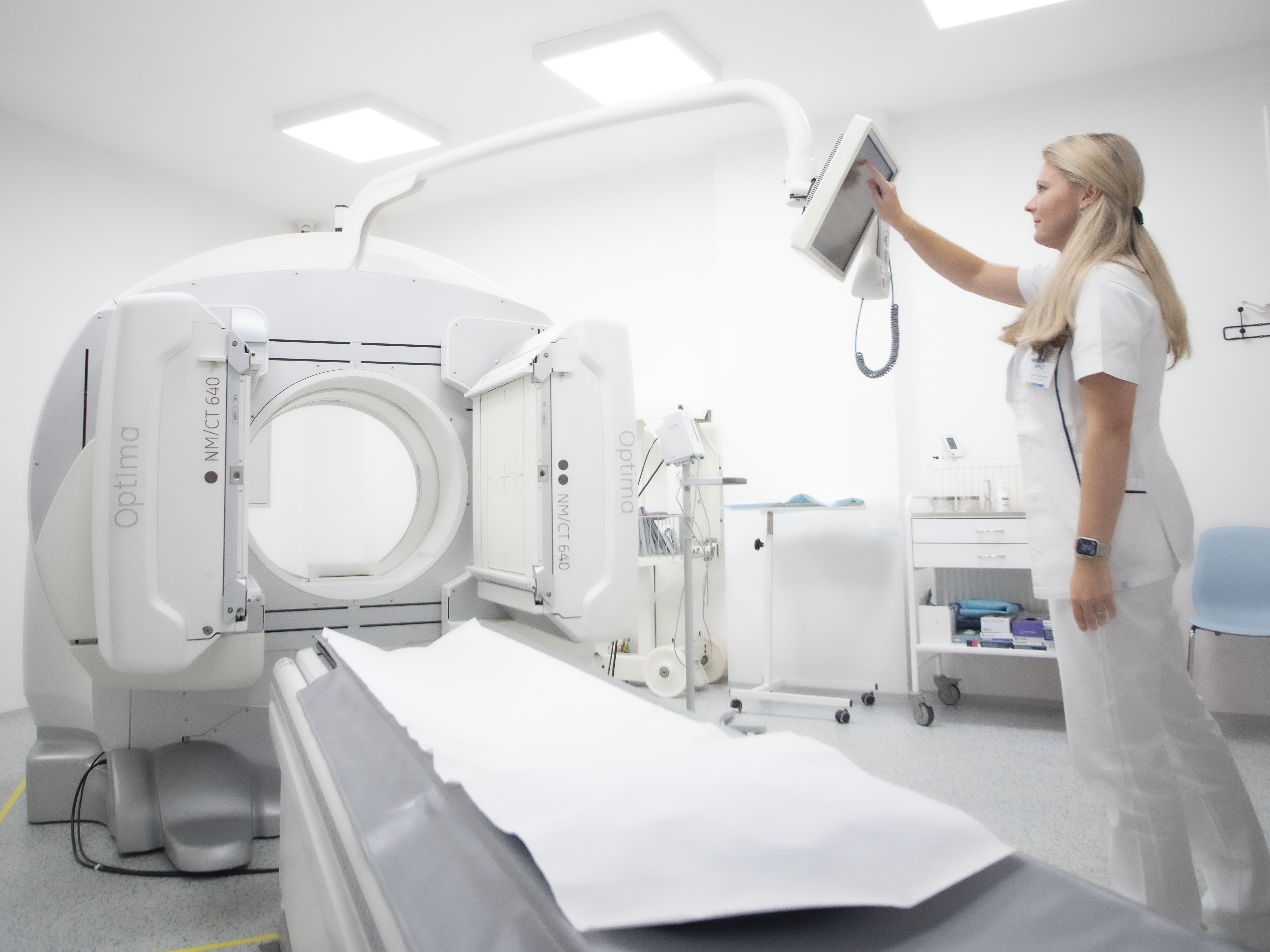
Principle and method
Radionuclide phlebography is a functional imaging method used in nuclear medicine to evaluate venous return and patency of the deep venous system of the lower limbs and pelvis.
After intravenous administration of a radiopharmaceutical into the venous system of the lower limbs (usually dorsally on the leg), its dynamic distribution in the proximal direction is monitored. The recording allows the speed, symmetry and continuity of flow in individual segments to be assessed.
Radiopharmaceutical: 99mTc-MAA (macroaggregate albumin) or 99mTc-colloid, which behaves passively and reflects the patency of the venous system, is most commonly used. The examination is performed using planar dynamic scintigraphy, sometimes supplemented with static images of the pelvis and abdomen.
Main clinical indications
- Diagnosis of deep vein thrombosis (DVT) – detection of venous obstruction, differentiation between acute thrombosis and chronic changes.
- Evaluation of venous collaterals – assessment of alternative drainage in case of obstruction.
- Follow-up after surgical or endovascular procedures – after phlebothrombolysis, stent placement or bypass.
- Chronic venous insufficiency – assessment of reflux and valve insufficiency.
- Differential diagnosis of lower limb oedema – differentiation between venous and lymphatic components (in combination with lymphoscintigraphy).
Contraindications and patient preparation
- Relative contraindications: pregnancy, allergy to the radiopharmaceutical used, severe local inflammation or infection at the injection site.
- Patient preparation: no special diet or fasting is required; moderate hydration is recommended.
Examination procedure
- Insertion of an intravenous cannula – usually into the dorsal vein of the leg.
- Administration of radiopharmaceutical (99mTc-MAA or 99mTc-colloid) in a dose of approximately 150–200 MBq.
- Dynamic scintigraphy – recording of the proximal progression of the radiopharmaceutical (blood flow) through the venous system of the lower limbs.
- Static images – pelvic, abdominal and chest areas as clinically required.
- Total examination time: 20–30 minutes.
Interpretation and clinical significance
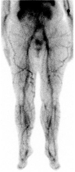
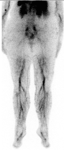
- Normal findings: symmetrical passage of the radiopharmaceutical through both lower limbs without signs of obstruction, rapid transport to the pelvic veins and vena cava.
- Acute HŽT: absence of flow in the section with thrombosis, or cessation of radiopharmaceutical progression.
- Chronic DVT: slowed flow, irregular distribution, presence of collaterals.
- Collateral circulation: visualisation of alternative venous pathways (paravertebral, superficial veins of the lower limbs).
The examination has the advantage of allowing functional assessment of flow dynamics and lower stress compared to conventional X-ray phlebography, as it does not use contrast agents.
Radiation exposure
- Effective dose: approx. 2–3 mSv.
- Radiopharmaceuticals are safe and well tolerated.
Summary for practice
Radionuclide phlebography is:
- A functional method for assessing venous patency and flow dynamics.
- Sensitive in the diagnosis of acute and chronic deep vein thrombosis.
- Useful for follow-up after surgical or endovascular procedures.
- Safe, with low radiation exposure and no risk of contrast nephropathy.
Although it is being partially replaced by ultrasound and CT, it remains a valuable alternative in situations where other methods are limited and complements the comprehensive diagnosis of venous diseases.
