Perfusion scintigraphy of the brain in epilepsy (ictal SPECT)
A practical guide for referring physicians
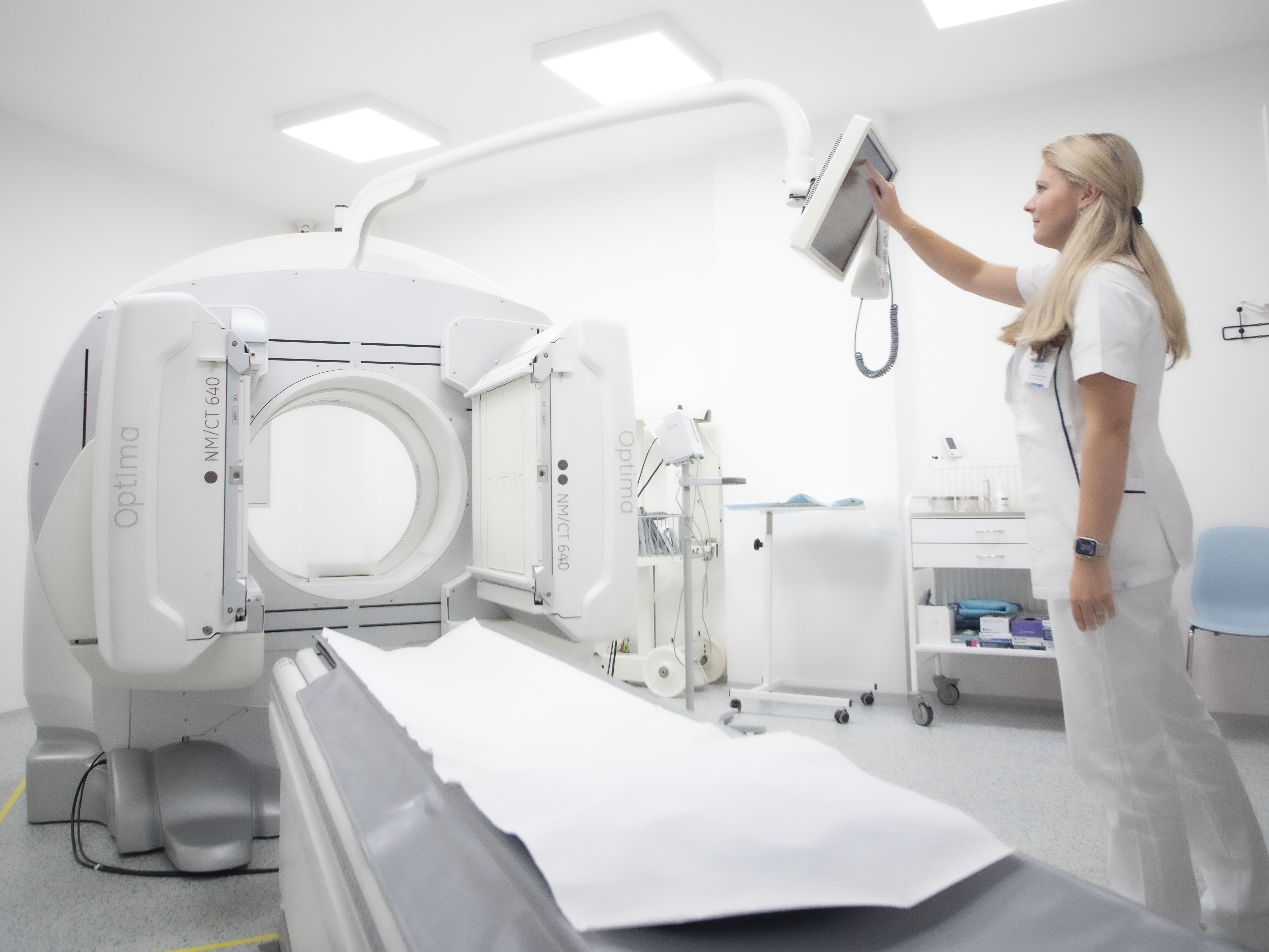
Principle and method
Perfusion scintigraphy of the brain using SPECT is a unique functional imaging method that allows for the evaluation of regional cerebral perfusion. In the ictal variant (performed during an epileptic seizure), it allows for the localisation of the epileptogenic focus in patients with drug-resistant epilepsy.
After intravenous administration of a lipophilic radiopharmaceutical (99mTc-HMPAO or 99mTc-ECD) during a seizure, it rapidly crosses the blood-brain barrier and is fixed in the brain tissue. The distribution of the radiopharmaceutical corresponds to the regional blood flow at the time of administration. Subsequent SPECT imaging, performed after the seizure has subsided, shows perfusion changes corresponding to ictal activity.
The method thus captures an "imprint" of blood flow during a seizure and is extremely valuable for localising the epileptic focus, especially in cases where EEG and MRI results are inconclusive.
Main clinical indications
- Drug-resistant epilepsy – determination of the epileptogenic zone prior to surgical treatment (especially temporal epilepsy).
- Differential diagnosis of seizure types – distinguishing between partial and generalised seizures.
- Complementary method in preoperative brain mapping – combination with EEG, MRI and PET.
- Research indications – study of the pathophysiology of epileptic seizures.
Interpretation and clinical significance
Typical ictal findings
- Hyperperfusion in the area of the epileptic focus at the time of the seizure.
- Perfusion enhancement is often greater than the epileptogenic cortex itself (due to the propagation of ictal activity).
Interictal findings
- Hypoperfusion in the epileptogenic focus outside of seizures.
- Interictal scintigraphy is used to supplement the comprehensive picture.
Highly valuable combination
- Comparison of ictal and interictal findings (known as SISCOM – Subtraction Ictal SPECT Coregistered to MRI) provides high accuracy in localising the epileptic focus and is one of the most advanced diagnostic methods in epileptology.
Practical information for the referring physician
- Patient preparation: hospitalisation in a specialised department with EEG monitoring; i.v. cannula inserted.
- Radiopharmaceuticals: 99mTc-HMPAO, 99mTc-ECD (lipophilic, fix in the brain shortly after administration).
- Examination procedure:
- continuous EEG monitoring of the patient,
- administration of radiopharmaceutical immediately after the onset of the seizure,
- SPECT imaging (even several hours after administration – distribution does not change),
- possibility of supplementing with an interictal study and correlation with MRI (SISCOM).
- Duration of the examination: imaging itself takes 30–60 minutes; logistics, including monitoring, take several hours.
- Radiation exposure: approx. 5–7 mSv.
- Contraindications: pregnancy; relative contraindications if safe EEG monitoring cannot be ensured.
Summary for practice
Perfusion scintigraphy of the brain in epilepsy (ictal SPECT) is:
- a unique functional method capable of localising the epileptogenic focus,
- key in the preoperative examination algorithm for drug-resistant epilepsy,
- highly effective in combination with EEG, MRI and PET (SISCOM),
- a method with high predictive value.
Correct indication and precise execution make ictal scintigraphy an essential diagnostic tool that can determine surgical treatment and lead to a significant improvement in the quality of life of patients with epilepsy.
