Sentinel lymph node scintigraphy
A practical guide for referring physicians
 Principle and method
Sentinel lymph node scintigraphy is a radionuclide examination designed to image the first lymph node (known as the sentinel node) to which lymph drains from the primary tumour. A radiopharmaceutical based on colloidal particles labelled with 99mTc is administered to the area surrounding the tumour. These particles are transported through the lymphatic system and captured in the sentinel node.
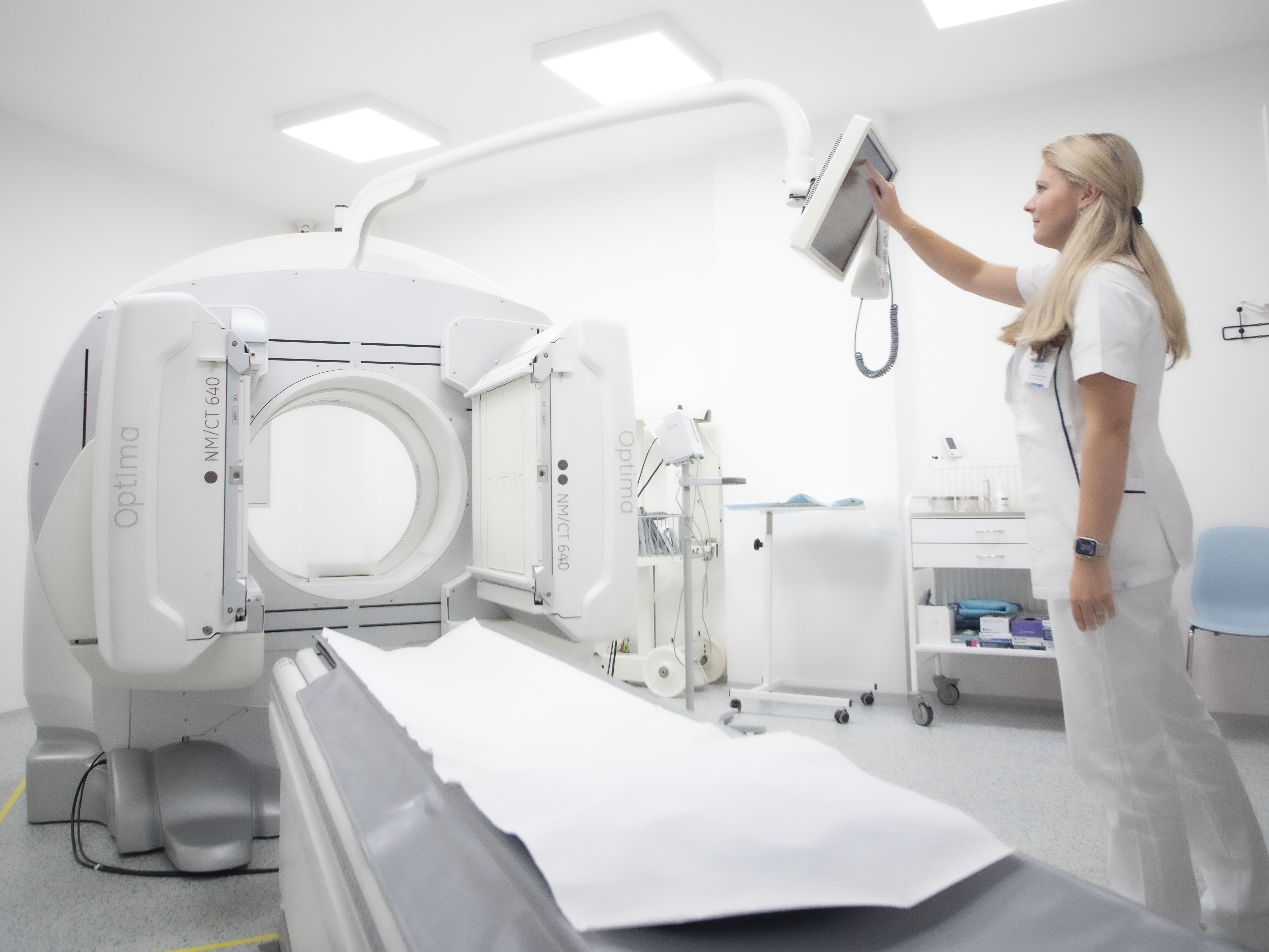
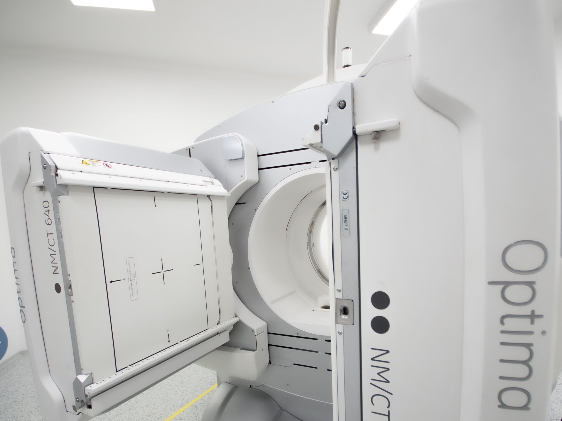
Planar scintigraphy and SPECT/CT enable precise localisation of the sentinel node, which is then used by the surgeon for targeted removal and histopathological examination. The method is minimally invasive, the burden on the patient is very low, and it significantly reduces the need for extensive lymphadenectomy.
Main clinical indications
- Breast cancer – standard part of staging
- Malignant melanoma – determination of regional lymph node involvement
- Carcinoma of the oral cavity, pharynx, vulva, penis – selected cases
- Other indications as determined by a multidisciplinary team
Interpretation
- The examination only determines the first node in the drainage – histology is necessary to confirm/rule out metastases
- Reasons for non-visualisation: lymphatic obstruction, atypical drainage, technical problems
Practical information for the referring physician
- Patient preparation: no preparation
- Examination duration: usually within one hour (includes administration of radiopharmaceutical, imaging)
- Radiation exposure: very low (<1 mSv)
- Contraindications: pregnancy – relative, must be considered on an individual basis
Summary for practice:
Sentinel lymph node scintigraphy is:
- a standard method in breast cancer and melanoma oncology,
- beneficial for precise surgical targeting and reduction of postoperative complications.
Correct indication and multidisciplinary team cooperation increase its clinical significance and benefit for the patient.
