Dynamic renal scintigraphy
A practical guide for referring physicians
Principle and method
Dynamic renal scintigraphy (nephrography) is a basic functional method in nuclear medicine for evaluating perfusion, function and urine outflow from both kidneys separately.
After intravenous administration of a radiopharmaceutical (most commonly 99mTc-MAG3 for evaluating tubular secretion or 99mTc-DTPA for glomerular filtration), its distribution and elimination are monitored in real time using dynamic scintigraphy.
The results are dynamic curves (nephrograms) that reflect:
- the blood supply to the kidney (perfusion phase),
- the accumulation of the radiopharmaceutical in the parenchyma (secretory phase),
- its excretion into the pelvis and ureter (excretory phase).
The examination thus provides comprehensive and quantitative information about both kidneys separately, including their relative function.
Main clinical indications
- Evaluation of differential renal function
- in congenital and acquired kidney diseases,
- before unilateral nephrectomy, surgical procedures or radiotherapy.
- Obstructive uropathy with furosemide administration
- evidence and quantification of the significance of urinary tract obstruction,
- assessment of the difference between obstruction and simple dilatation (hydronephrosis vs. retention).
- Congenital developmental defects
- Reflux nephropathy, duplication of the ureter and renal pelvis, renal ectopia.
- Arterial hypertension
- in combination with pharmacological intervention (captopril test) to demonstrate renovascular hypertension.
- Acute and chronic kidney disease
- assessment of functional reserve and renal clearance.
- Postoperative and post-traumatic conditions
- monitoring of transplanted kidney function,
- follow-up after urinary tract reconstruction procedures.
Interpretation
- Normal findings:
- rapid onset of perfusion, accumulation in the parenchyma and gradual excretion into the pelvis and ureter,
- the curve has a typical shape with a rapid rise and fall.
- Pathological findings:
- obstruction – accumulation of radiopharmaceutical in the pelvis without subsequent elimination (so-called "obstructive renogram"),
- parenchymal involvement – reduced accumulation and flat, slow curve progression,
- vascular disorder – delayed perfusion phase.
Supplemented with pharmacological tests:
- Furosemide test (F+): administration of furosemide in cases of suspected obstruction → assessment of whether the pelvis empties.
- Captopril test: see chapter on renovascular hypertension.
Practical information for the referring physician
- Patient preparation:
- sufficient hydration (0.5–1 litre of fluids 30–60 minutes before the examination),
- empty bladder,
- Examination procedure:
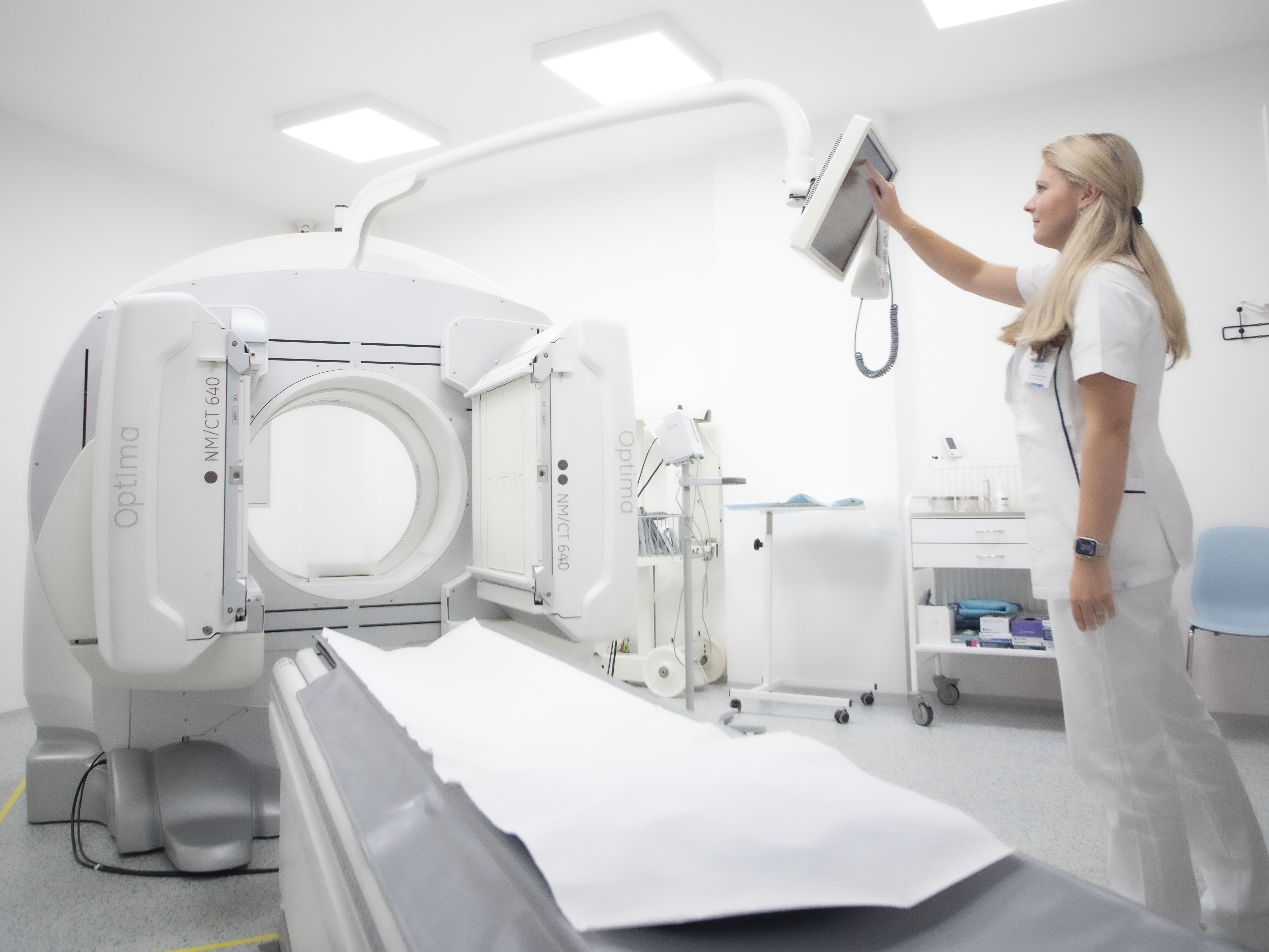
- Intravenous administration of radiopharmaceutical,
- dynamic imaging takes 30 minutes, patient lies on their back
- administration of furosemide (diuretic test) or enalapril (RVH provocation) as needed.
- Radiopharmaceuticals:
- 99mTc-MAG3 (most common),
- 99mTc-DTPA (GFR assessment),
- Radiation exposure: low, less than 1 mWv.
- Contraindications: pregnancy; relative in patients unable to cooperate.
Summary for practice
Dynamic renal scintigraphy is:
- the basic method for functional assessment of the kidneys and urinary tract,
- indispensable in the diagnosis of obstructive uropathies,
- valuable in determining differential renal function prior to surgical procedures,
- useful in the diagnosis of renovascular hypertension and monitoring of transplanted kidneys,
- safe, accessible and low in radiation exposure.
Proper indication and combination with pharmacological tests increase its informative value and significantly contribute to decisions on further procedures in nephrology and urology.
