Static renal scintigraphy (99mTc-DMSA)
Practical guide for referring physicians
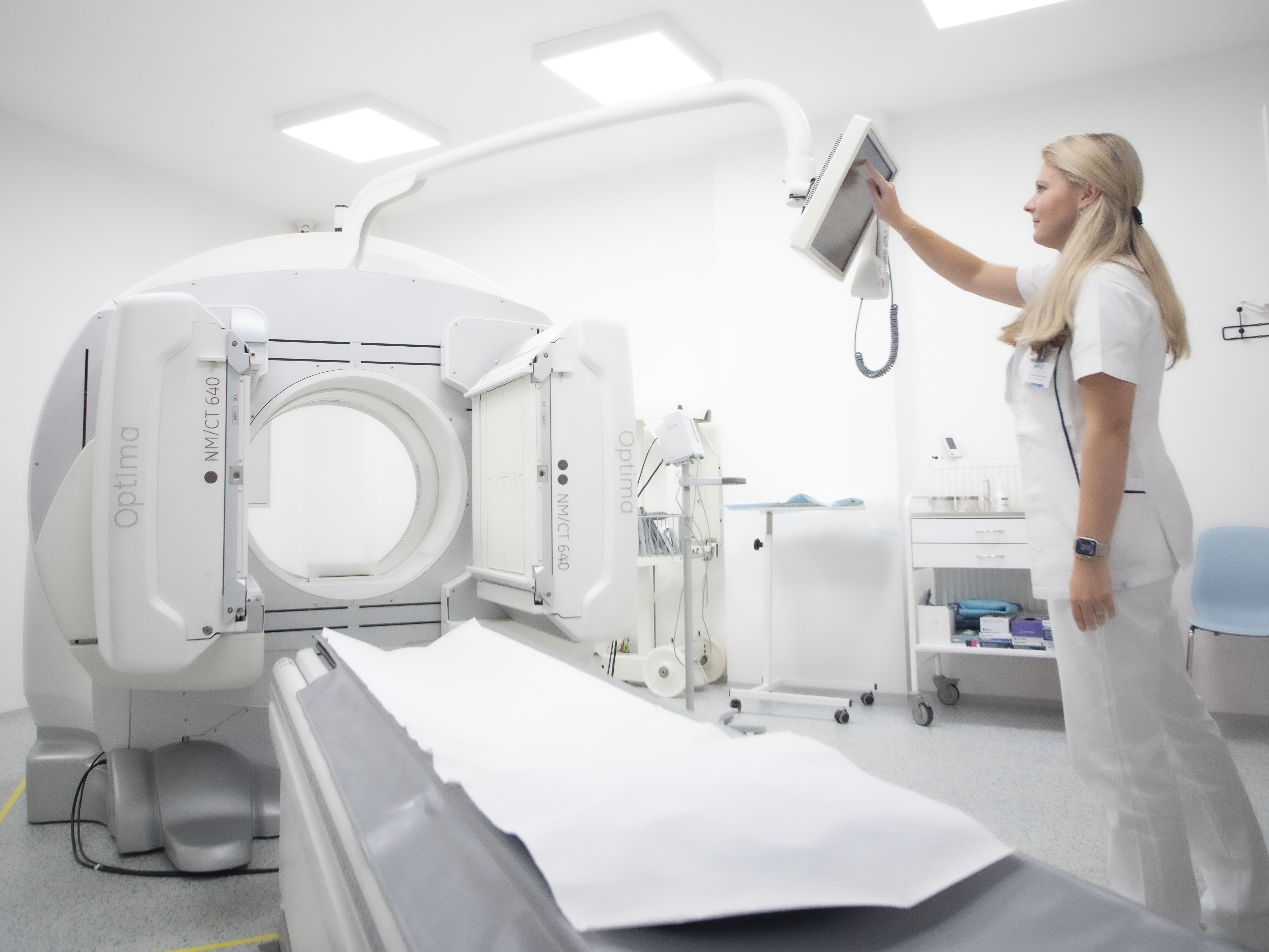
1. Principle and method of examination
Static renal scintigraphy using the radiopharmaceutical 99mTc-DMSA (dimercaptosuccinic acid) is a radionuclide imaging method designed for detailed evaluation of cortical functional tissue in the kidneys.
After intravenous administration, DMSA selectively binds to the proximal tubules of the renal cortex, where it is retained for a long time. Thanks to this mechanism, the examination provides a high-quality image of the morphology and functional integrity of the renal parenchyma with high spatial resolution.
Unlike dynamic renal examinations (MAG3, DTPA), the aim is not to evaluate flow or drainage, but to provide a static image of functional tissue, enabling the identification of focal defects, scars, hypoplasia and other structural changes.
The method is considered the gold standard for assessing cortical kidney involvement, especially in paediatric nephrology, but it also has wide application in adult patients.
2. Main clinical indications
Static renal scintigraphy is of fundamental importance in the following clinical situations:
2.1 Infectious kidney disease
- Acute pyelonephritis – evidence of focal parenchymal involvement, especially in atypical or complicated cases
- Chronic pyelonephritis – detection of permanent cortical scars and assessment of their extent
2.2 Reflux nephropathy
- Assessment of the consequences of vesicoureteral reflux, especially in paediatric patients
- Detection and quantification of renal scars that cannot be reliably visualised by other methods
2.3 Congenital renal malformations
- Renal ectopia
- Hypoplasia or aplasia
- Duplication of the renal system
- Asymmetry in kidney size and function
2.4 Differential diagnosis of focal changes
- Distinguishing functional renal tissue from non-functional structures (scars, cysts, post-operative changes)
- Clarification of findings in unclear lesions detected by ultrasound, CT or MR
2.5 Quantification of relative renal function
- Determination of the relative functional contribution of individual kidneys, particularly important before surgical procedures or during long-term monitoring of chronic nephropathies
3. Interpretation of findings
3.1 Normal findings
- Symmetrical size and shape of both kidneys
- Homogeneous, uniform accumulation of radiopharmaceutical in the cortical layer
- No focal uptake defects
3.2 Pathological findings
- Focal accumulation defects corresponding to scars, inflammatory changes or ischaemic damage
- Diffuse reduction in uptake in global parenchymal involvement
- Asymmetry in size, shape or function of the kidneys
- Reduced relative functional contribution of one kidney
The examination allows for a semi-quantitative assessment of the relative function of individual kidneys, which is clinically significant in monitoring disease progression and deciding on therapeutic procedures.
4. Practical information for the referring physician
- Patient preparation:
No special preparation is necessary, fasting is not required - Hydration: Adequate hydration
is recommended before and after administration of the radiopharmaceutical - Radiopharmaceutical administration:
Intravenous administration of 99mTc-DMSA - Time interval until imaging:
Optimally 2–4 hours after administration, according to the facility protocol - Duration of examination:
Total time spent by the patient approx. 3–4 hours, imaging itself approx. 15–30 minutes - Radiation exposure:
Low (usually 1–2 mSv), significantly lower than for CT examinations
The method is also suitable for paediatric patients - Contraindications:
- Pregnancy (relative)
- Breastfeeding – recommended to stop breastfeeding for 24 hours after administration
Summary for practice
Renal scintigraphy with captopril test is:
- a sensitive functional method for detecting renovascular hypertension,
- capable of distinguishing whether angiographic stenosis is haemodynamically significant,
- useful in selecting patients for revascularisation procedures,
- safe, simple and less costly than morphological imaging methods.
Correct indication and correlation with clinical and angiographic findings significantly increase its value in the diagnosis and management of patients with severe arterial hypertension.
