Lymphoscintigraphy for limb swelling: How it works and when it helps detect lymphoedema
Why do limbs swell and when is lymphoedema the cause?
Swelling of the lower or upper limbs is a very common problem. Sometimes it is a normal reaction to prolonged standing, travelling or venous problems.
But if the swelling:
- persists for a long time,
- worsens during the day,
- is stiff or tense,
- appears after surgery or radiation therapy,
it may be caused by lymphoedema — a disorder of lymph drainage.
Lymph is a clear fluid that removes excess water, proteins and waste products from tissues. If the lymphatic vessels or nodes are damaged or overloaded, fluid begins to accumulate → swelling occurs.
This is where lymphoscintigraphy plays a crucial role — an examination that can accurately show how lymph flows in the limb.
What is lymphoscintigraphy?
Lymphoscintigraphy is a safe and minimally invasive nuclear medicine examination. A small dose of
radiopharmaceutical is used to monitor:
- the speed of lymph flow,
- the function of the lymphatic pathways and nodes,
- the location of any slowdowns or blockages,
- the presence of collateral pathways,
- dermal backflow — the return of lymph to the tissues, which is typical of advanced lymphoedema.
The examination uses the radiopharmaceutical 99mTc-nanocolloid, which is administered by a very gentle injection under the skin of the hand or foot. The
radiation exposure is very low (less than a standard CT scan) and therefore safe even for long-term patient monitoring.
When is lymphoscintigraphy recommended?
The examination is most often indicated:
1️⃣ For swelling after surgery or radiation
E.g. after breast surgery, gynaecological procedures, lymph node removal or radiotherapy.
2️⃣ When primary (congenital) lymphoedema is suspected
Often in younger people, sometimes from puberty.
3️⃣ For chronic swelling without a clear cause
When ultrasound of the veins shows no disorder, but the oedema persists.
4️⃣ To distinguish between lymphoedema and lipoedema
Lipedema (painful fat deposits on the thighs/arms) is treated differently than lymphedema.
5️⃣ After repeated infections (e.g. erysipelas)
Inflammation can damage the delicate lymphatic vessels.
How does the examination work?
The examination takes two hours, but the patient has breaks between the individual steps.
The injection and imaging are painless.
1. Gentle injection
The radiopharmaceutical is administered subcutaneously (under the skin), usually between the toes. The dose
administered is very small and harmless. The
entire procedure takes only a few seconds and the burning sensation is minimal. This is followed by imaging, which takes two minutes, after which the patient lies on a bed for 30 minutes.
2. Imaging after rest
Cameras monitor how the radiopharmaceutical begins to be absorbed into the lymphatic vessels. This step takes approximately 20 minutes.
3. Active phase
To support lymph flow, the patient:
- walks for 30 minutes in the case of the lower limbs,
- repeatedly clenches and unclenches their fist if the upper limbs are being examined.
This part mimics natural movement, which helps the lymph flow.
This is followed by another scan after exercise lasting approximately 15 minutes.
What can the examination reveal?
1️⃣ Slowed lymph flow
Common in early lymphedema, which is still clinically barely visible.
2️⃣ Complete blockage of the lymphatic pathway
Typical after lymph node removal or large scars.
3️⃣ Collateral (alternative) pathways
The body looks for a "detour" so that the lymph can continue to flow.
4️⃣ Dermal reflux
Lymph returns to the soft tissues → causing stiff, chronic swelling.
5️⃣ Insufficient lymph node function
For example, after damage caused by radiation.
Is the examination safe?
Yes. The radiation dose is very low — for example, with an application of 50 MBq, it is less than 1 mSv.
For comparison: a standard chest CT scan has a 7–8 times higher dose.
Contraindications:
- pregnancy (only in exceptional cases),
- allergy to products containing human albumin,
- breastfeeding requires a 13-hour break and restriction of close contact with the child (recommended by the National Radiology Standard).
How will the results help the patient?
Lymphoscintigraphy results help to:
- confirm or rule out lymphoedema,
- determine the appropriate intensity of lymphatic drainage and compression therapy,
- set up a long-term physiotherapy plan,
- prevent complications such as worsening swelling or infection,
- evaluate the success of treatment.
A correct diagnosis is key to relieving the patient's symptoms and controlling the swelling.
Conclusion: Lymphoscintigraphy provides certainty where other methods fall short
Ultrasound shows veins, but not the lymphatic system. CT and MRI show structures, but not lymph flow.
Lymphoscintigraphy is the only method that can directly and accurately monitor lymph flow in the limbs and detect even subtle disorders.
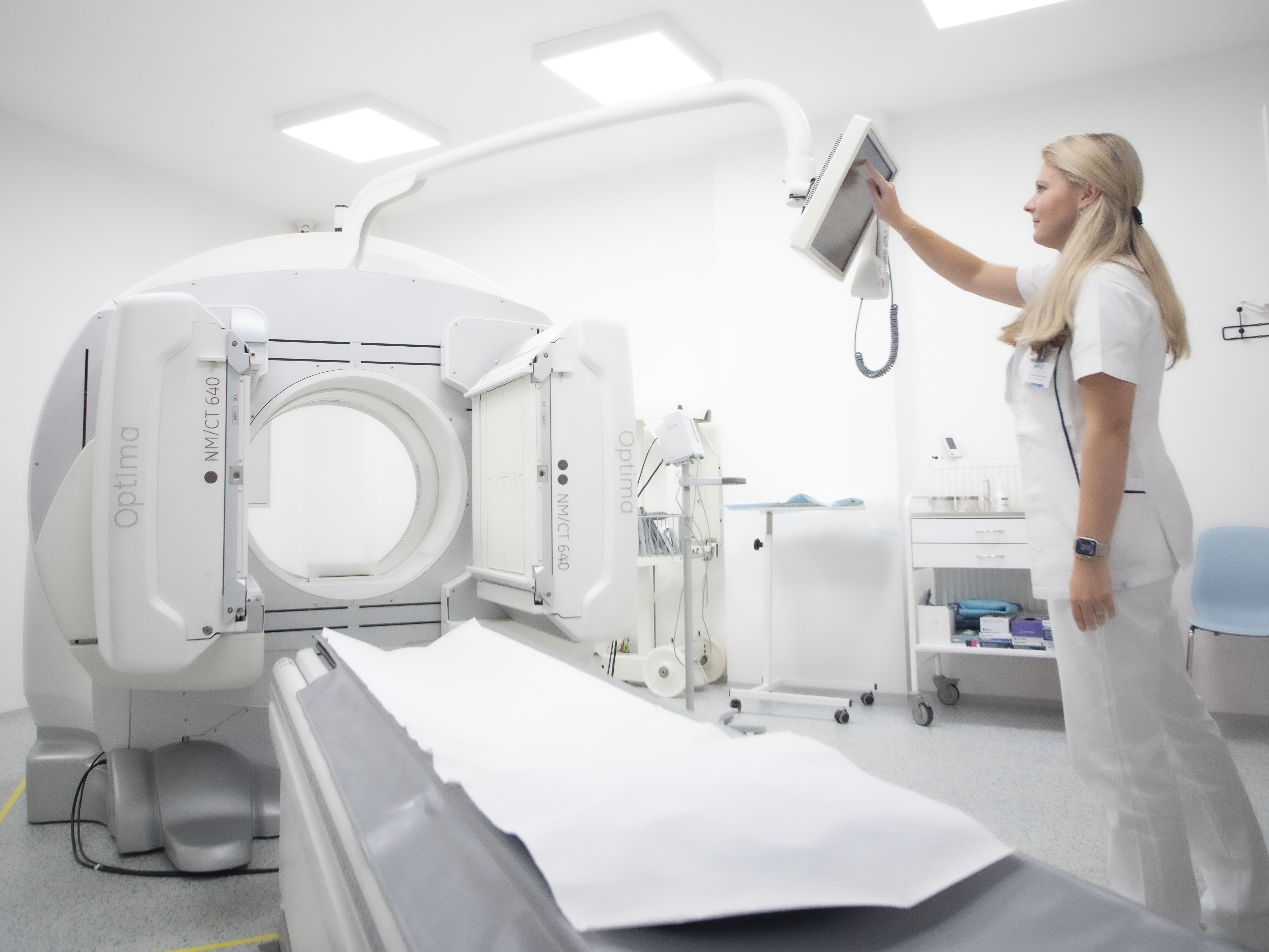
At PMCD, we perform lymphoscintigraphy according to strict standards and using modern cameras, which allows us to evaluate even very early or atypical forms of lymphoedema.
